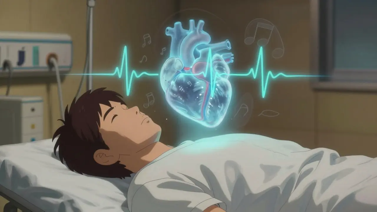
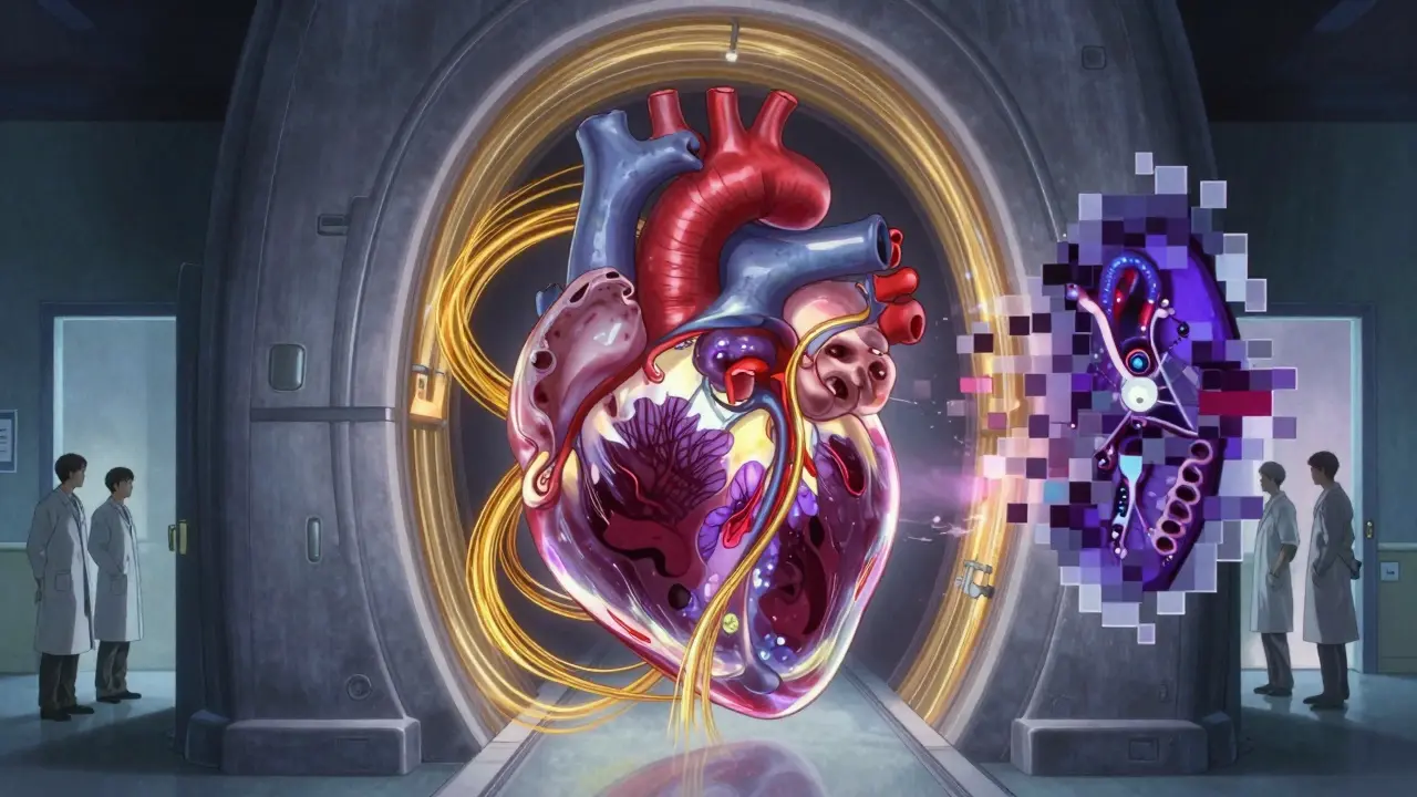
When your doctor suspects something’s off with your heart, you’ll likely hear about two tests: cardiac MRI and echocardiography. Both show your heart in detail, but they’re not the same. One uses sound waves. The other uses magnets. One is fast and cheap. The other is slower but more precise. Knowing the difference isn’t just about understanding medical jargon-it can change how your treatment unfolds.
Echocardiography, or "echo," has been the go-to heart test for decades. It uses high-frequency sound waves, like sonar, to make moving pictures of your heart. A probe placed on your chest sends out pulses, and the echoes that bounce back create real-time video. No radiation. No needles. Just a gel on your skin and a technician moving a device around.
It’s quick-usually under 30 minutes. It’s portable. You can get an echo in an ambulance, a doctor’s office, or even at your bedside in the hospital. That’s why about 15 million echocardiograms are done every year in the U.S. alone. It’s the first step for almost everyone: checking for valve leaks, measuring how well your heart pumps, or spotting fluid around the heart.
Standard echo numbers? Normal left ventricular ejection fraction (LVEF) is 50-75%. That’s the percentage of blood your heart pushes out with each beat. Normal left ventricular end-diastolic dimension (LVEDD)? 37-56 mm. Interventricular septal thickness? 6-11 mm. These values help doctors decide if your heart’s thickened, enlarged, or weak.
But echo has limits. If you’re overweight, have lung disease, or your ribs block the view, the images get fuzzy. That’s called a "poor acoustic window." In those cases, echo might miss early signs of disease. One cardiologist in Birmingham told me last year, "I’ve had patients where echo showed normal function, but MRI found scar tissue-changes that were already affecting their risk of arrhythmia."
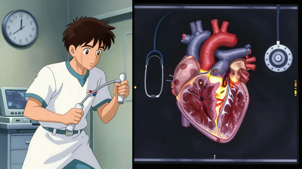
Cardiac MRI is like a high-res 3D scan of your heart. It uses strong magnetic fields and radio waves-not sound-to create images. It doesn’t rely on how well sound travels through your chest. It sees everything: the shape of each chamber, the thickness of every wall, and even the texture of the heart muscle itself.
That last part matters. MRI can detect scar tissue, inflammation, or fat deposits inside the heart muscle. This is done with a technique called late gadolinium enhancement (LGE). When a patient has suspected myocarditis or heart damage from chemotherapy, MRI is often the only test that confirms it. A 2023 study in JACC: CardioOncology found that 2D echo underestimated ejection fraction by 3% on average-and in 10% of cancer patients, that error meant misclassifying who was at risk for heart damage.
For volume measurements, MRI is the gold standard. Unlike echo, which guesses heart shape using math formulas, MRI counts every drop of blood in the heart using 3D imaging. No assumptions. Just direct measurement. Studies show echo consistently reports smaller heart chambers and thicker walls than MRI. One 2011 study found echo overestimated septal thickness by 1.1 mm on average. That might not sound like much, but in a patient with hypertrophic cardiomyopathy, it could mean missing a dangerous diagnosis.
Accuracy matters most when tracking changes over time. A 2022 paper in the Journal of the American College of Cardiology showed MRI’s inter-observer variability for ejection fraction was just 2.6%. Echo? 6.8%. That means two different doctors reading the same echo could disagree by nearly 5 percentage points. With MRI, they’d be within 1%.
Here’s the real-world breakdown:
One key advantage of echo is right ventricular function. In patients with pulmonary hypertension or congenital heart disease, echo’s myocardial performance index (MPI) is often more reliable than MRI for detecting severely reduced right heart function. MRI can struggle with the thin, fast-moving walls of the right ventricle.
But MRI wins in tissue detail. If you’ve had a heart attack and your echo says "mildly reduced function," but you’re still getting chest pain and fatigue, MRI might show small areas of scar tissue that echo missed. That changes your treatment-maybe you need a different medication, or a device like an ICD.
Let’s talk money and time. An echocardiogram costs between $500 and $1,500. A cardiac MRI? $1,500 to $3,500. That’s why most hospitals use echo first. But cost isn’t just the test price-it’s the whole chain.
A 2022 JAMA Internal Medicine analysis found that while MRI costs $2,000 more upfront, it often prevents unnecessary follow-up tests. In complex cases, patients who got MRI after an unclear echo saved money overall-about $750 per patient-by avoiding extra scans, hospital visits, or misdiagnoses.
But access? That’s the real hurdle. In community hospitals, 78% have echo available immediately. Only 35% offer cardiac MRI within a week. In the UK, wait times for non-urgent cardiac MRI can stretch beyond 14 days. One Reddit user, CardiacTech2023, wrote: "I’ve seen patients wait six weeks for MRI while their symptoms worsened. Echo gave us something to act on right away."
Performing echo takes skill, but it’s easier to learn. The American Society of Echocardiography says you need 300-500 supervised scans to become proficient. For cardiac MRI? 1,000-1,500. That’s why fewer radiologists and cardiologists are fully trained in MRI interpretation.
But technology is closing the gap. New ultrasound machines like Philips’ EPIQ CVx now use AI to auto-calculate ejection fraction, reducing variability to just 4.2%. Meanwhile, Siemens’ new 0.55T MRI machine, launched in June 2023, can scan patients with pacemakers and defibrillators-something older MRIs couldn’t touch.
Emerging techniques like parametric mapping (T1, T2, ECV) let MRI measure tissue properties without contrast dye. This could make it safer and more useful for kidney patients or those allergic to gadolinium.
The future? Hybrid protocols. By 2030, experts predict combining real-time echo with detailed MRI tissue maps will become standard for complex cardiomyopathies. Imagine an echo guiding a catheter during a procedure, then an MRI confirming the tissue changes afterward. That’s the next step.
If you’re scheduled for one of these tests, ask:
Don’t assume echo is "good enough." If your symptoms don’t match your echo results, push for further imaging. Many patients are told, "Your echo looks fine," when MRI later reveals early disease. One 2023 study found that 19% of patients with suspected heart failure had normal echo but abnormal MRI findings.
And if you have an implanted device-pacemaker, ICD, or loop recorder-don’t assume MRI is off-limits. New low-field machines are changing that. Always ask: "Is there a safe way to do an MRI with my device?"
Yes, for accuracy and consistency. Cardiac MRI measures heart volume and ejection fraction without guessing the heart’s shape, unlike echocardiography, which relies on geometric formulas. Studies show MRI has far less variability between doctors-just 2.6% compared to echo’s 6.8%. It’s the gold standard for tracking changes over time, especially in conditions like heart failure or cardiomyopathy.
No. Echocardiography shows structure and movement, but it can’t tell the difference between healthy muscle and scar tissue. That’s where cardiac MRI excels. Using late gadolinium enhancement (LGE), MRI can pinpoint areas of fibrosis or scarring from past heart attacks, inflammation, or genetic conditions-something echo simply can’t see.
If the echo images were unclear-due to body type, lung disease, or poor windows-or if the results didn’t match your symptoms. For example, if you have fatigue and abnormal ECG but normal echo, MRI might reveal hidden scar tissue, inflammation, or early thickening of the heart muscle. It’s also used for diagnosing conditions like myocarditis, sarcoidosis, or amyloidosis that echo often misses.
The main risk is from gadolinium contrast, which can rarely cause kidney issues or a skin condition called nephrogenic systemic fibrosis in patients with severe kidney disease. Newer contrast agents are safer, but screening is still done. Also, older pacemakers or metal implants may not be MRI-safe-but newer low-field MRI machines (like 0.55T) can now scan most patients with devices. Always tell your provider about any implants.
The scan itself takes 45-60 minutes. But interpretation is complex and requires specialized training. In most hospitals, results take 3-7 days. In busy centers or community hospitals without dedicated cardiac MRI experts, it can take two weeks or more. Echocardiography results are often available the same day.
It depends. Older pacemakers and defibrillators are still a contraindication for standard 1.5T or 3T MRI machines. But since June 2023, new 0.55T MRI systems have been approved for use with many previously incompatible devices. Always check with your cardiologist and the imaging center-some devices are now labeled "MRI-conditional" and can be scanned safely under specific conditions.
Cardiac MRI is more accurate. A 2023 study in JACC: CardioOncology found 2D echo underestimated ejection fraction by a median of 3% compared to MRI, with variations as wide as ±15%. That’s enough to misclassify patients in critical situations, like cancer patients at risk for heart damage. MRI’s accuracy comes from 3D volumetric measurements without geometric assumptions.
Echocardiography is your heart’s first look. Fast. Widely available. Good for most situations. Cardiac MRI is the deep dive. Slower. More expensive. But when precision matters-when scar tissue, inflammation, or subtle changes could change your future-it’s unmatched. They’re not rivals. They’re partners. One opens the door. The other walks you through the whole house.

Look, I’ve seen too many patients get told "your echo looks fine" and then wind up in the ER six months later with a scarred-up heart. MRI isn’t just fancy-it’s life-saving when echo misses the subtle stuff. I don’t care if it costs more or takes longer. If your symptoms don’t match the echo, push back. Hard. 🤝

While echocardiography remains indispensable for its accessibility and real-time utility, cardiac magnetic resonance imaging offers unparalleled accuracy in tissue characterization and volumetric quantification. The inter-observer variability data cited-2.6% for MRI versus 6.8% for echo-is statistically significant and clinically meaningful, particularly in longitudinal monitoring of cardiomyopathies. Furthermore, late gadolinium enhancement remains the non-invasive gold standard for myocardial fibrosis detection.

Echo first. MRI if it doesn’t add up. Simple.

YES. This. 🙌 I had a "normal echo" after months of fatigue and chest tightness. My cardiologist said "maybe anxiety." Six weeks later, I got an MRI and found patchy fibrosis from silent myocarditis. They missed it. I almost missed it too. If your body’s screaming and the echo says "all good," keep pushing. You deserve better. 💪❤️

So let me get this straight-we spend $3,000 on a machine that takes an hour, just because echo sometimes can’t see through fat? I mean, I get it, but also… is this really how we’re fixing healthcare? Next thing you know, we’ll be doing PET scans for sprained ankles.

My mom’s echo came back "borderline" after her pneumonia. She was told to wait and see. But she kept feeling off-dizzy, short of breath even sitting down. We pushed for an MRI. Turned out she had early amyloidosis. Echo would’ve never caught it. I’m so glad we didn’t take "it’s probably fine" as an answer. If you’re not feeling right, don’t let cost or convenience silence you.

Ugh, I hate when docs treat MRI like it’s the holy grail. It’s not magic. It’s just a different lens. I had a friend who got an MRI because her echo was "inconclusive"-turned out she just had a weird chest wall anatomy. No disease. No treatment needed. Just a fancy scan that scared her for three weeks. Sometimes echo is enough. Stop overmedicalizing everything.

I’ve been a nurse for 18 years, and I’ve seen echo save lives every day. It’s quick. It’s safe. It’s everywhere. MRI? Beautiful tech. But not everyone needs it. If your doctor says echo is enough, trust them. If you’re scared, ask: "What’s the next step if this doesn’t explain my symptoms?" That’s the right question. Not "can I get an MRI?"

Let’s be real: MRI is overused. It’s expensive. It’s slow. It’s full of false positives. And don’t get me started on gadolinium-some patients develop chronic pain from it. Meanwhile, echo has improved dramatically. AI-assisted measurements? Down to 4.2% variability. That’s almost MRI-level. Why are we still pushing MRI like it’s the only way? Because hospitals make more money off it. And patients? They’re just being sold fear.