TL;DR
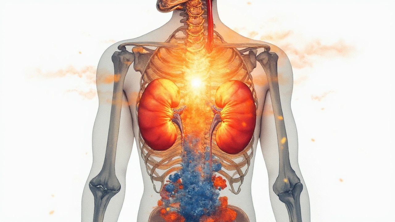
If your kidneys are struggling, your bones feel it. Healthy kidneys activate vitamin D (they convert 25‑OH vitamin D to 1,25‑OH₂ vitamin D, also called calcitriol). Calcitriol helps you absorb calcium from food. When kidneys are damaged, that activation drops, phosphate builds up, and your body releases more PTH to keep calcium stable. This bundle-abnormal calcium, phosphate, PTH, and vitamin D-is called CKD‑mineral and bone disorder (CKD‑MBD).
Early on, your calcium might look “normal” because PTH compensates. As CKD progresses or vitamin D runs low, calcium can tip under the lab range. High phosphate doesn’t help-it binds calcium and lowers the free (ionised) part that nerves and muscles rely on.
What you feel ranges from subtle to dramatic. The quiet stuff: tingling lips or fingertips, muscle cramps, twitchy eyelids, brittle nails, and bone aches. More obvious red flags: carpopedal spasms (hands locking), facial twitching, hoarse voice or stridor (throat muscles tightening), and seizures. If spasms, breathlessness, or seizures hit, that’s an emergency.
Before changing treatment, make sure the number is real. In the UK, labs usually report calcium in mmol/L alongside albumin. Low albumin makes total calcium look lower than it truly is. Many clinics correct total calcium for albumin using: corrected Ca (mmol/L) = measured Ca + 0.02 × (40 − albumin g/L). If you’re borderline or symptomatic, an ionised calcium is more reliable.
Other causes can push calcium down in kidney disease too:
Reading the pattern helps you and your team target the fix. Here’s a quick lab logic you can apply with your clinician:
| Scenario | Calcium | Phosphate | PTH | Likely driver |
|---|---|---|---|---|
| CKD secondary hyperparathyroidism | Low to low‑normal | High | High | Low calcitriol + phosphate retention |
| Hypoparathyroidism | Low | High | Low/normal (inappropriate) | PTH lack (postsurgical/autoimmune) |
| Hungry bone syndrome | Low | Low/normal | High | Rapid skeletal uptake after PTH drop |
| Vitamin D deficiency (non‑dialysis) | Low | Normal/low | High | Poor intake/absorption or low sun |
| Medication‑related (e.g., cinacalcet) | Low | Variable | Lowered by drug | Oversuppressed PTH/calcium shift |
Ranges vary by lab, but as a rough UK guide: corrected calcium 2.2-2.6 mmol/L; phosphate 0.8-1.5 mmol/L; PTH depends on assay (often ~1.6-6.9 pmol/L in people with healthy kidneys; higher targets may be acceptable in dialysis). eGFR tells you the CKD stage and how often to monitor.
Evidence you can lean on: KDIGO CKD‑MBD guidance stresses treating trends, not single numbers, and avoiding calcium/phosphate overload. UK NICE guidance recommends vitamin D repletion in deficiency for CKD and tailored use of active vitamin D only when indicated. The UK Kidney Association echoes limiting calcium‑based binders when phosphate is hard to control.

Start with a tidy checklist and move step by step. You’ll save time and avoid bouncing between fixes that fight each other.
| CKD Stage / Situation | Monitoring Frequency (Ca/Phos) | PTH Monitoring | When to Act |
|---|---|---|---|
| Stage 3 (eGFR 30-59) | Every 6-12 months | Once, then by trend | Correct vitamin D deficiency; investigate persistent low Ca or rising PTH |
| Stage 4 (eGFR 15-29) | Every 3-6 months | Every 6-12 months | Consider active vitamin D if high PTH + low Ca after basics fixed |
| Stage 5 not on dialysis | Every 1-3 months | Every 3-6 months | Tighten phosphate control; careful with calcium-based binders |
| Dialysis (HD/PD) | Monthly | Every 3-6 months | Adjust binders, dialysate calcium, and active vitamin D as needed |
| Post-parathyroidectomy | Weekly initially | As advised by surgeon | Watch for hungry bone-may need higher calcium + calcitriol short term |
Targets are nuanced. Many UK renal teams aim to keep corrected calcium in the lab’s normal range, phosphate in or near normal (especially in non‑dialysis CKD), and PTH “appropriate for CKD stage” rather than forcing it to the range seen in healthy kidneys. The thread running through KDIGO guidance: avoid long stretches of high calcium and high phosphate together.
Simple home checklist you can actually use:

Sometimes examples land better than theory. Here are three that map to common clinic stories in the UK.
Scenario 1: Stage 3 CKD, fatigue, and mild hypocalcemia. Your corrected calcium is 2.15 mmol/L, phosphate 1.1 mmol/L, PTH slightly high, 25‑OH vitamin D is low. You start vitamin D loading as per GP guidance, tweak breakfast (swap processed cereals for porridge without phosphate additives), add a daily walk, and recheck in 6 weeks. Calcium moves to 2.25 mmol/L, PTH drifts down, symptoms fade. No binders needed. Lesson: fix vitamin D and food first when phosphate isn’t high.
Scenario 2: Dialysis with muscle cramps and low post‑dialysis calcium. You’re already on sevelamer. Calcium runs 2.05-2.15 mmol/L with tingling after sessions. The unit reviews binders, nudges dialysate calcium up within the safe range, and adds a small dose of alfacalcidol. Symptoms settle; phosphate holds steady. Lesson: sometimes it’s a dialysate and dosing dance-small changes add up.
Scenario 3: After parathyroid surgery, calcium crashes. You feel shaky; calcium is 1.85 mmol/L, phosphate is 0.7 mmol/L, PTH is low. The team diagnoses hungry bone syndrome and uses IV calcium briefly, then oral calcium plus calcitriol with close monitoring. Over weeks, bones “fill up,” labs stabilise, and supplements taper. Lesson: timing matters-expect short bursts of higher support after surgery.
Food swaps that protect bone and heart without making meals boring:
Mini‑FAQ
Next steps and troubleshooting
When to seek urgent help (don’t wait for a routine clinic slot):
Pro tips I share with patients in clinic:
Behind the scenes, your team is balancing two risks: the misery of low calcium and the long‑term harm from high calcium plus high phosphate. The sweet spot is personal and may change over time. Stay curious, keep your numbers handy, and nudge your plan when life (or labs) changes.
Key references used by UK renal teams: KDIGO CKD‑MBD updates (2017 and subsequent commentaries), NICE guidance on CKD and vitamin D, UK Kidney Association phosphate binder recommendations, and National Kidney Foundation clinical resources. If a recommendation here affects your treatment, check it with your clinician who knows your history and local lab targets.
Oh wonderful, another deep dive into CKD‑MBD-because we clearly don’t have enough checklists to fill our lives, right? Still, the reminder to correct calcium for albumin is gold; many miss that and chase a phantom hypocalcemia. If you’re already on phosphate binders, double‑check the calcium load-they can tip you over the edge. And that little tidbit about citrate in dialysate? It explains the occasional twitch without any new meds. Keep an eye on magnesium; it’s the sneaky side‑kick that can block PTH and keep calcium low. Remember, the “safe” target isn’t a prison‑cell number, it’s a range that keeps vessels from calcifying. TL;DR: confirm, don’t guess, and treat the whole mineral bundle, not just calcium alone.
Hey folks, just wanted to add a friendly nudge: staying on top of your labs every few weeks can really prevent those scary spasms from sneaking up. A quick phone call to your clinic for the latest phosphate and PTH can save you a trip later. Keep a simple log of symptoms-tingling, cramps, even subtle fatigue-so you have something concrete to discuss. You’ve got this, and the more proactive you are, the smoother the management will be.
From a physiological standpoint, the interplay between reduced 1,25‑OH₂ vitamin D synthesis and phosphate retention creates a feedback loop that perpetuates secondary hyperparathyroidism. When the kidneys fail to hydroxylate 25‑OH vitamin D, intestinal calcium absorption drops, prompting the parathyroids to secrete more PTH, which in turn attempts to mobilise calcium from bone. This compensatory mechanism, while initially protective, eventually leads to bone demineralisation and vascular calcification if left unchecked. Therefore, a comprehensive assessment-including ionised calcium, magnesium, and vitamin D metabolites-is essential before initiating any therapeutic adjustments.
Allow me to elaborate at length, for those who enjoy a thorough exposition on the intricacies of CKD‑related hypocalcemia. First, it is paramount to recognise that the kidneys serve not merely as a filtration organ but also as a pivotal site for the activation of vitamin D, converting 25‑hydroxyvitamin D into the biologically active 1,25‑dihydroxy form, calcitriol, which in turn facilitates intestinal calcium absorption. When renal function declines, this conversion wanes, precipitating a cascade of metabolic disturbances. Simultaneously, phosphate excretion is impaired, leading to hyperphosphatemia; the elevated phosphate binds free calcium, diminishing the ionised fraction that is physiologically active. The parathyroid glands, sensing this deficit, ramp up parathyroid hormone (PTH) secretion in a compensatory effort to maintain calcium homeostasis, a state known as secondary hyperparathyroidism. However, chronic elevation of PTH exerts deleterious effects on bone turnover, promoting resorption that further destabilises skeletal integrity. Moreover, the high‑phosphate, low‑calcium milieu fosters vascular calcification, a leading cause of cardiovascular morbidity in dialysis patients. Therapeutically, the clinician must adopt a multipronged strategy: judicious use of calcium‑based phosphate binders, careful supplementation with active vitamin D analogues such as calcitriol or alfacalcidol, and, when appropriate, calcimimetics like cinacalcet, albeit with vigilant monitoring to avoid oversuppression of calcium levels. Dietary phosphate restriction, though challenging, remains a cornerstone of management, complemented by regular dialysis sessions calibrated to optimise phosphate clearance. Importantly, serum magnesium should not be overlooked, as hypomagnesemia can blunt PTH responsiveness and exacerbate hypocalcaemia. Finally, the frequency of laboratory monitoring must be tailored to the CKD stage, with more intensive surveillance in advanced disease to promptly detect and correct metabolic derangements before they translate into clinical sequelae. In sum, an integrated approach that addresses the intertwined pathways of vitamin D metabolism, phosphate balance, PTH dynamics, and magnesium status offers the best prospect for stabilising calcium levels while mitigating the long‑term complications of CKD‑MBD.
Great post! 😊, remember to check your calcium *and* phosphate levels, because they love to play hide‑and‑seek, especially on dialysis days, 😅, and don’t forget magnesium – it’s the unsung hero, 😇, keep a medication list handy, it saves time, and always ask your nephrologist about vitamin D dosing, 📋.
One could argue that the entire emphasis on aggressive calcium supplementation is a misguided relic of outdated protocols that ignore the nuanced risk of vascular calcification and the emerging data supporting conservative management strategies which, if not reconsidered, may inadvertently exacerbate morbidity in the CKD population.
Vitamin D is essential.
Keep the optimism flowing; regular monitoring and a balanced diet can dramatically improve quality of life, especially when you pair low‑phosphate meals with appropriate vitamin D supplementation; you’re on the right track, keep it up!
In many cultures, traditional foods like fermented soy or low‑phosphate broths naturally support mineral balance; incorporating such staples can be a gentle, culturally resonant way to aid CKD management without relying solely on pharmaceuticals.
Frankly, the article glosses over the fact that many clinicians still prescribe calcium‑based binders indiscriminately, despite substantial evidence linking them to accelerated arterial calcification; this oversight can mislead patients into a false sense of security.
The drama of a sudden seizure from hypocalcemia is something straight out of a medical thriller, yet it underscores the sheer volatility of mineral imbalances in CKD; without vigilant care, the stakes are literally life‑or‑death.
I appreciate how the guide consolidates lab interpretation steps; having a clear decision tree for calcium, phosphate, and PTH really streamlines multidisciplinary discussions and reduces unnecessary testing.
Thanks for the thorough overview; I’ll definitely share this checklist with my clinic’s nursing staff so we can all stay aligned on monitoring frequencies and treatment thresholds.
One must consider that the pharmaceutical industry may be subtly influencing guideline committees; after all, the push for newer active vitamin D analogues coincides conveniently with their profit motives, raising questions about the objectivity of the recommendations presented herein;
From a grammatical standpoint, the article maintains commendable clarity, yet it could benefit from more precise usage of terms such as "hypocalcaemia" versus "hypocalcemia" to adhere to standardized medical nomenclature across British and American publications.
Excellent exposition! 😊; however, one minor point: the phrase "calcium‑based binders when phosphate is hard to control" could be refined for greater syntactic accuracy, perhaps by rephrasing to "calcium‑based binders are less advisable when phosphate control proves challenging".
Building on the earlier note about active vitamin D analogues, it’s crucial to titrate calcitriol cautiously in dialysis patients, starting low and adjusting based on ionised calcium trends; too aggressive a dose can precipitate hypercalcemia and worsen vascular calcification.
Exactly! Also, don’t forget to educate patients on the timing of phosphate binders relative to meals-taking them with food maximises efficacy, and pairing with a small calcium supplement post‑dialysis can smooth out the calcium dip you often see overnight.